The Planetary Society, dedicated to exploring the solar system and seeking life beyond Earth, asked Michael S. Gazzaniga what a human community will look like on Mars.
Talking with the world’s leading expert on cognitive neuroscience, one of the field’s founders, and one of the greatest minds of our time, the Society wanted to know Gazzaniga’s opinions in relation to the human brain. In other words, will the brain change? Look different structurally or perform functionally differently? In other words, will the brain enable the mind on Mars differently than on Earth?
Gazzaniga’s response, as reported on the Society’s website, tells us something about Gazziniga’s 45 years of research studying the most important organ in the human body:
“New environments are fascinating and challenging. We combine elements of the new place with different outcomes. Yet, the piece of biologic tissue that does these wonderful things is the same for all humans and hasn’t changed in thousands of years. On Sunday afternoon on Mars, citizens of the New City will still love a cold beer and a good NFL game.”
What makes each of us uniquely human fascinates cognitive neuroscientists such as Gazzaniga. But before they dissect the cognitive processes that contribute to our uniqueness, they study the basics of brain structure. In other words, the journey of unraveling the mind-brain connection begins with brain anatomy.
The following description provides a general overview of the brain’s structure.
Different Regions of the Brain
The Cerebral Cortex
This is the outer covering of the brain, the one with all the bumps (gryi or the singular gyrus) and folds (sulci or the singular sulcus). The cortex has a large number of connections – 75% in total. The other 25% of connections link to other brain regions, such as the nervous system.
Six layers of nerve cells and the pathways connecting these layers comprise the cerebral cortex.
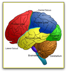
Scientists subdivide the cerebral cortex into four lobes:
- Frontal lobe
- Parietal lobe
- Temporal lobe
- Occipital lobe
There are two brain hemispheres, the left and right brain hemispheres. Hence, scientists say that there are actually eight lobes, referring, for example, to the lobes as plural (two frontal lobes, two parietal lobes, etc.)
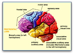
Scientists localize some functions to particular lobes. For instance, speech production or generation is generally thought to occur in the left frontal lobe. However, molecular and cellular brain research from the last decade has shown that many particular “functions” are actually more widespread in one or more brain lobes or regions, and that neuronal circuits connect these distributed functional areas.
The Frontal Lobes
The frontal lobes are more highly developed in humans than other animals and are the centers for many higher functioning aspects of behavior, such as language, planning, generating ideas, problem solving, working memory, and personality. The frontal lobes are the last part of the brain to grow to full size, with full maturity occurring late into adolescence.
The primary motor cortex called the M1, an area associated with planning, executing, and refining motor movements, is located in the posterior area of the frontal lobes.
The prefrontal cortex
The prefrontal cortex is the anterior area (foremost area) of the frontal lobe. It differs from other parts of the frontal cortex by having an additional layer of neurons. This area is thought to control complex cognitive behaviors, such as modeling correct social behavior, determining consequences of certain behaviors, working toward goals, and predicting outcomes.
The Parietal Lobes
A strip or groove of cortex running across the top of the brain called the central sulcus separates the frontal lobes from the parietal lobes, which are located posterior to (behind) the frontal lobes.
The somatosensation strip or the S1 is the first area or strip of the parietal lobe, a strip of cortex that contains a map of the entire body for processing touch sensations. After the S1 strip, areas of the parietal lobes mediate the integration of sensory information, connecting touch with visual data or memory.
Visuospatial processing, and some aspects of language and attention are also conducted in the parietal lobes. An area of the left parietal lobe also manages motor control.
The Temporal Lobes
The anterior or front part of the temporal lobes are bounded by the frontal lobe to one side, and the brain stem to the other. The upper part of the brain stem called the pons actually connects and extends from the temporal lobes. To the rear of the temporal lobes are the parietal and occipital lobes.
The temporal lobes are further segmented into three gyri: the superior or upper; the medial or mid; and the inferior or lower.
An area of the superior gyrus is the primary auditory control center, also known as Heschl’s gyrus, that receives auditory inputs from the ears. On the left side of the primary auditory center, recognition of language sounds takes place. On the right side, recognition of non-verbal sounds such as tone, rhythm, and emotion takes place.
Parts of the inferior temporal lobes contribute to the recognition of specific visual stimuli, such as faces, objects, and animals. Scientists think that the inferior temporal lobes store memories in a highly organized way.
The Occipital Lobes
Lying below the parietal and temporal lobes are the occipital lobes – the smallest of the four lobes. They are located at the back or rearmost part of the brain. These lobes contain the roadmap for visual processing.
An area called Brodmann area 17 or V1 is the primary visual cortex, residing in the medial (mid) part of he occipital lobe. This area extends to the posterior pole of the lobe. The V1 is also referred to as the striate cortex because it contains a large stripe of myelin. Areas located around the striate are called extrastriate cortex, and they mediate the perception of forms, movement, movement direction, location, and color discrimination.
Wider Areas of Brain Structure
The regions described above provide an overview of the brain as if visually examined from the outside. But there are also many areas inside the brain responsible for memory, perception, learning, emotions, and other cognitive systems. Most of the deep brain structures connect with multiple brain areas and regions.
Scientists use another categorization system to name large sections of the brain, calling these sections the forebrain, midbrain, and hindbrain.
The Forebrain
The forebrain contains the cerebral cortex, and the basal ganglia. The basal ganglia are clusters of nuclei consisting of several interrelated structures that scientists believe are involved with motor control and movement. The amygdala is one component of the basal ganglia.
The forebrain also contains the limbic system, often referred to as the brain’s emotional center, controlling emotions such as fear, anger, and pleasure. The limbic system includes the hippocampus, which plays an important role in long-term memory and spatial memory. It also includes the mammillary bodies, anterior thalamic nucleus, cingulate cortex, the formix, and the entorhinal cortex.
The limbic works with both higher cortical brain structures as well as those of the brain stem.
The Thalamus
The thalamus, responsible for relaying sensation, spatial sense and motor signals, is centrally located on top of the brainstem. It helps regulate consciousness, sleep, and alertness. Some textbooks on the brain place the thalamus in the forebrain, others place it in the midbrain, still others place it between the forebrain and midbrain.
The Midbrain
The midbrain’s structures are involved with the processing of auditory and visual reflexes, such as turning the head toward a sound or moving the eyes toward a visual stimulus. These structures are called the tectum or colliculi.
The Hindbrain
This area of the brain contains structures outside of the cerebral cortex: the pons, medulla oblongata, and cerebellum. The spinal cord becomes the medulla oblongata as it makes its way into the cranium. This main nervous system connector carries messages between the spinal cord and brain. It also controls basic bodily functions such as respiration, heart rate, and digestive activities.
The pons lies right above the medulla, relaying signals from the medulla to the brain. It also links the cerebellum to the rest of the brain.
The cerebellum lies on the dorsal (back) part of the brainstem at about ear level. This structure controls balance, contributes to the learning of coordinated movements, and processes sensory information used by motor systems.
Brainy Careers
Those who make a living studying the brain describe it as a passion – not a job. For those interested in probing how the brain enables the mind, or in other words, how the mind-brain connection makes humans unique among all living creatures, several career paths are possible.
An bachelor’s or graduate degree in psychology provides an excellent background for those interested in the brain. Most research positions require a Ph.D. Contact psychology schools for more information.
Careers that Require a Strong Background in Psychology:
- Cognitive Neuropsychologist
- Cognitive Neuroscientist
- Development Cognitive Neuropsychologist
- University teaching and research
- School Neuropsychology Consultation
- Neuropsychology researcher for public and private organizations
- Neurogenetics Psychologist
- Neuropathology Specialist
- Behavioral Neuropsychologist
- Behavioral Neuroscientist
- Forensic Neuropsychologist
- Rehabilitation Neuropsychologist
- Dementia Neuropsychologist